LAG-3 Negatively Regulates T Cell Functions
LAG-3 Interacts with Multiple Ligands and Inhibits T cell Responses
Lymphocyte Activation Gene 3 (LAG-3), also known as CD223, is a type I transmembrane protein that belongs to the immunoglobulin superfamily (IgSF). It is expressed as a dimer or as an oligomer on the surface of activated CD4+ and CD8+ T cells, regulatory T cells, Tr1 cells, natural killer cells, and plasmacytoid dendritic cells (pDCs).1, 2 Similar to PD-1 and CTLA-4, LAG-3 is also up-regulated on exhausted T cells in cancer.2,3 The LAG-3 protein is structurally homologous to CD4 in that it has four extracellular Ig-like domains, but it also contains an extra loop on the membrane-distal Ig-like domain that is not present in CD4. This extra loop allows the LAG-3 protein to bind to MHC class II molecules expressed on antigen-presenting cells (APCs) or tumor cells with significantly higher affinity than CD4, and negatively regulate T cell receptor (TCR) signaling.4, 5
In addition to MHC class II molecules, Galectin-3, LSECtin, and Fibrinogen-like protein 1 (FGL1) are also LAG-3 ligands.2, 6-8 Significantly, all three of these proteins are expressed on various tumor cell types and inhibit T cell responses, making it unclear which ligands are primarily responsible for the immunosuppressive functions of LAG-3.6, 8 Although the mechanisms by which LAG-3 signals are not well understood, LAG-3 has been shown to negatively regulate T cell activation, proliferation, and cytokine production, inhibit pDC activation, and enhance the suppressive activity of regulatory T cells.9-15 LAG-3 has also recently been suggested to inhibit cytokine secretion by mature natural killer cells.16 On regulatory T cells, LAG-3 was found to engage MHC class II on dendritic cells (DCs) and inhibit DC maturation and their immunostimulatory capacity .17 Conversely, a soluble variant of LAG- 3 was shown to bind to MHC class II molecules on immature DCs and induce their maturation, leading to enhanced CD8+ T cell antigen cross-presentation.18, 19
Similar to PD-1, LAG-3 is frequently up-regulated on tumor-infiltrating lymphocytes (TILs) and while blockade of LAG-3 alone was found to be only weakly effective in reducing tumor growth in some mouse tumor models, blockade of both LAG-3 and PD-1 synergistically enhanced anti-tumor immunity and survival, when compared with anti-PD-1 alone.20, 21 In many of these mouse tumor models, the improvement in the anti-tumor immune response was attributed to an increase in the number of tumor-infiltrating CD8+, or CD8+ and CD4+ T cells, and an accompanying increase in IFN-gamma production that was observed following anti-LAG-3/anti-PD-1 treatment. As a result, researchers are now evaluating an antagonistic anti-LAG-3 antibody either alone, or in combination with anti-PD-1, in clinical trials.22, 23 Additionally, due to the observation that soluble LAG-3 has immune-stimulatory effects, a soluble recombinant form of LAG-3, known as IMP321, is also being evaluated in phase I testing in combination with anti-PD-1.
Bio-Techne offers R&D SystemsTM bioactive recombinant proteins for LAG-3 and its ligands, along with fluorochrome-conjugated antibodies for detecting LAG-3, Galectin-3, and LSECtin to further our understanding of the immunomodulatory effects of LAG-3.
LAG-3 and LAG-3 Ligands - Products by Molecule
LAG-3 Binds to MHC II, Galectin-3, FGL1, and LSECtin and Suppresses T Cell Activity

LAG-3 is an immunosuppressive receptor that inhibits T cell activity and promotes the suppressive activity of regulatory T cells. LAG-3 binds to multiple ligands including MHC class II, Galectin-3, LSECtin, and Fibrinogen-like protein 1 (FGL1), which have all been reported to have T cell inhibitory effects. Similar to PD-1, TIM-3, TIGIT, and BTLA, LAG-3 is also up-regulated on exhausted T cells and natural killer cells in cancer and is thought to contribute to their dysfunction (left). On regulatory T cells, LAG-3 expression enhances their suppressive function and can inhibit dendritic cell maturation and immunostimulatory capacity through its interaction with MHC class II (right).
Analysis of the Binding Activity of R&D Systems Recombinant Human LAG-3 and Hepassocin/FGL1 Proteins
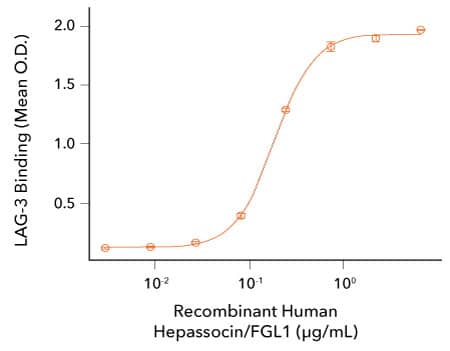
LAG-3 Binds to Hepassocin/FGL-1. Recombinant Human LAG-3 Fc Chimera (R&D Systems, Catalog # 2319-L3) was coated on a plate at 1 ug/mL and the indicated concentrations of Recombinant Human Hepassocin/FGL1 His tag (R&D Systems, Catalog # 10285-HE) were added. The concentration of Recombinant Human Hepassocin/FGL1 that produces 50% of the optimal binding response is 0.05-0.4 ug/mL.
Assessment of the Bioactivity of R&D Systems Recombinant Cynomolgus Monkey LAG-3 Protein

LAG-3 Induces TNF-alpha Secretion by Mouse Immature Dendritic Cells. JAWSII mouse immature dendritic cells were treated with the indicated concentrations of Recombinant Cynomolgus Monkey LAG-3 (R&D Systems, Catalog # 9992-L3). TNF-alpha secretion was measured in cell culture supernatants using the Mouse TNF-alpha Quantikine™ ELISA Kit (R&D Systems, Catalog # MTA00B). The ED50for this effect is 0.15-0.9 ug/mL in the presence of a cross-linking antibody, Mouse Anti-His Tag Monoclonal Antibody (R&D Systems, Catalog # MAB050R).
-
He, Y. et al. (2016) Lymphocyte-activation gene-3, an important immune checkpoint in cancer. Cancer Sci. 107:1193. PMID: 27297395.
-
Anderson, A.C. et al. (2016) Lag-3, Tim-3, and TIGIT: Co-inhibitory receptors with specialized functions in immune regulation. Immunity 44:989. PMID: 27192565.
-
Wherry, E.J. (2001) T cell exhaustion. Nat. Immunol. 12:492. PMID: 21739672.
-
Huard, B. et al. (1994) Lymphocyte-activation gene 3/major histocompatibility complex class II interaction modulates the antigenic response of CD4+ T lymphocytes. Eur. J. Immunol. 24:3216. PMID: 7805750.
-
Huard, B. et al. (1995) CD4/major histocompatibility complex class II interaction analyzed with CD4- and lymphocyte activation gene-3 (LAG-3)-Ig fusion proteins. Eur. J. Immunol. 25:2718. PMID: 7589152.
-
Xu, F. et al. (2014) LSECtin expressed on melanoma cells promotes tumor progression by inhibiting antitumor T-cell responses. Cancer Res. 74:3418. PMID: 24769443.
-
Kouo, T. et al. (2015) Galectin-3 shapes antitumor immune responses by suppressing CD8+ T cells via LAG-3 and inhibiting expansion of plasmacytoid dendritic cells. Cancer Immunol. Res. 3:412. PMID: 25691328.
-
Wang, J. et al. (2019) Fibrinogen-like protein 1 is a major immune inhibitory ligand of LAG-3. Cell 176:334. PMID: 30580966.
-
Workman, C.J. et al. (2009) LAG-3 regulates plasmacytoid dendritic cell homeostasis J. Immunol. 182:1885. PMID: 19201841.
-
Huard, B. et al. (1996) T cell major histocompatibility complex class II molecules down-regulate CD4+ T cell clone responses following LAG-3 binding. Eur. J. Immunol. 26:1180. PMID: 8647185.
-
Baixeras, E. et al. (1992) Characterization of the lymphocyte activation gene 3-encoded protein. A new ligand for human leukocyte antigen class II antigens. J. Exp, Med. 176:327. PMID: 1380059.
-
Hannier, S. et al. (1998) CD3/TCR complex-associated lymphocyte activation gene-3 molecules inhibit CD3/TCR signaling. J. Immunol. 161:4058. PMID: 9780176.
-
Huang, C.T. et al. (2004) Role of LAG-3 in regulatory T cells. Immunity 21:503. PMID: 15485628.
-
Workman, C.J. & D.A. Vignali (2003) The CD4-related molecule, LAG-3 (CD223), regulates the expansion of activated T cells. Eur. J. Immunol. 33:970. PMID: 12672063.
-
Workman, C.J. et al. (2004) Lymphocyte activation gene-3 (CD223) regulates the size of the expanding T cell population following antigen activation in vivo. J. Immunol. 172:5450. PMID: 15100286.
-
Narayanan, S. et al. (2020) LAG-3 is a central regulator of NK cell cytokine production.
-
Liang, B. et al. (2008) Regulatory T cells inhibit dendritic cells by lymphocyte activation gene-3 engagement of MHC class II. J. Immunol. 180:5916. PMID: 18424711.
-
Triebel, F. (2003) LAG-3: a regulator of T-cell and DC responses and its use in therapeutic vaccination. Trends Immunol. 24:619. PMID: 14644131.
-
Casati, C. et al. (2006) Soluble human LAG-3 molecule amplifies the in vitro generation of type 1 tumor-specific immunity. Cancer Res. 66:4450. PMID: 16618772.
-
Woo, S.R. et al. (2012) Immune inhibitory molecules LAG-3 and PD-1 synergistically regulate T-cell function to promote tumoral immune escape. Cancer Res. 72:917. PMID: 22186141.
-
Andrews, L.P. et al. (2017) LAG3 (CD223) as a cancer immunotherapy target. Immunol. Rev. 276:80. PMID: 28258692.
-
Dempke, W.C.M. et al. (2017) Second- and third-generation drugs for immuno-oncology treatment - the more the better? Eur. J. Cancer 74:55. PMID: 28335888.
-
Marin-Acevedo, J.A. et al. (2018) Next generation of immune checkpoint therapy in cancer: new developments and challenges. J. Hematol. Oncol. 11:39. PMID: 29544515.