Replace Western Blot Stripping and Reprobing with Automated RePlex Assays on Jess and Abby
Run Two Immunoassays in the Same Capillary
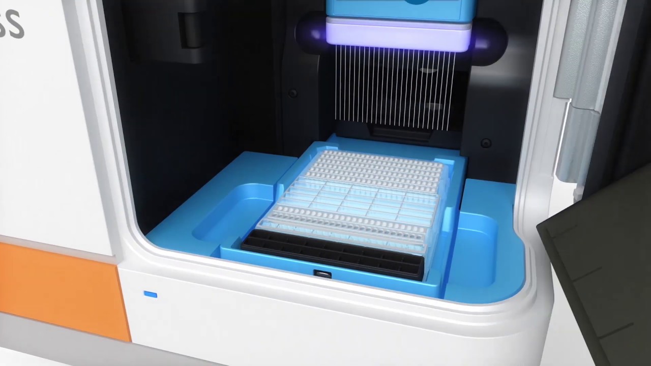
With the new RePlex™ feature on Jess™ and Abby™, you can run two Simple Western™ immunoassays within the same capillary to get all your rich protein characterization data from just one sample! Watch the video to see RePlex in action on Jess.
- Quantify expressed phosphorylated target and total target levels
- Normalize with total protein detection in the same capillary
- Greater flexibility in immunoprobe selection
- More data points per sample
- Save time and money on consumables
RePlex - A Strip and Reprobe Western Blot Alternative
RePlex enables two-step immunoassays that rely on the removal of antibodies from the first round of probing (Probe 1) prior to a second round of probing with fresh antibodies (Probe 2). Signal intensity and reproducibility are not compromised between Probe 1 and Probe 2. With RePlex on Simple Western, you'll never have to deal with Western blot stripping and reprobing again!
Reproducible Results Between Immunoprobe Cycles
Lane view in Compass for Simple Western™ shows similar band intensities and quantitation shows comparable peak areas for AKT1 and AKT2 in each probing cycle of a RePlex.

Choose the Probing Order with RePlex
Regardless of the order in which each protein was probed, AKT1 and AKT2 in MCF7 lysates detected in Probe 1 or Probe 2 in a RePlex assay show excellent reproducibility and similar signal intensity across both probing cycles of a RePlex. Protein integrity and re-probing are not compromised by antibody removal in RePlex assays.

Efficient Antibody Removal Between Probing Cycles
Because the samples are covalently bound to the capillary wall, RePlex completely and reproducibly removes antibodies between cycles without loss of signal intensity.
Lane view in Compass for Simple Western shows immunoassay signal for Probe 1 and no residual signal for Probe 2 for multiple targets in Jurkat and MCF7 cell lines.

Immunoassay and Total Protein Detection in the Same Capillary
The second cycle of RePlex can be dedicated to total protein detection so that you can normalize your data with confidence. All the steps of RePlex are automatically performed with Jess and Abby, providing more data, and lowering the cost of reagents and consumables per result.
Automated Total Protein Normalization
The figure shows AKT phosphorylation in MCF7 lysates untreated and activated with h-IGF1. (A-B) Phospho-AKT and pan AKT were detected in Probe 1 using chemiluminescence and NIR fluorescence, respectively, while total protein signal was detected in Probe 2. (C) The Peak Table in Compass for Simple Western shows automated normalization of phosphorylated and pan AKT signal to total protein signal, demonstrating quantitation of target protein expression.
