TNF Superfamily Ligands and Receptors

TNF Superfamily Ligand-Receptor Interactions & Select Functions
TNF Superfamily Proteins
The tumor necrosis factor superfamily (TNFSF) in humans consists of 19 proteins that play important roles in regulating the proliferation, differentiation, activation, and survival of multiple immune and non-immune cell types. Some TNF superfamily proteins transduce signals that lead to apoptosis, while others are involved in regulating innate and adaptive immune responses, and still others promote cell-type specific responses such as osteoclast development. TNF-alpha was the first TNF superfamily protein to be identified, followed by Lymphotoxin-alpha (LT-alpha)/TNF-beta, which was found to bind to the same receptor as TNF-alpha and share 50% amino acid sequence homology. Seventeen other related proteins were subsequently identified based on their sequence homologies with TNF-alpha. With the exception of Lymphotoxin-alpha (LT-alpha)/TNF-beta, all members of the TNF superfamily are type II transmembrane proteins that share a common structural motif known as the TNF homology domain (THD), which mediates both their trimerization and receptor-binding capabilities. While most TNF superfamily proteins are synthesized as membrane-bound proteins, cleavage of their extracellular domains by specific metalloproteinases gives rise to soluble forms of the proteins. TNF superfamily proteins are primarily expressed by antigen-presenting cells, such as dendritic cells, macrophages, B cells, and neutrophils, but they can also be expressed by T cells, natural killer cells, basophils, eosinophils, mast cells, endothelial cells, and thymic epithelial cells.
TNF Receptor Superfamily
Members of the TNF superfamily bind to receptors belonging to the TNF receptor superfamily (TNFRSF), which consists of 29 receptors in humans and 32 in mice. With the exception of BAFF R, BCMA, TACI, and XEDAR, which are type III transmembrane proteins, TNF superfamily receptors are type I transmembrane proteins or soluble decoy receptors. The extracellular domains of all TNF receptors are characterized by the presence of one or more highly conserved, cysteine-rich domain(s) that mediates ligand binding. High affinity binding of a TNFSF trimer to its receptor promotes receptor clustering and trimerization, leading to formation of a 3:3 hexameric ligand-receptor complex and activation of downstream signaling pathways. As is outlined in the graphic above, several TNF superfamily ligands can bind to more than one receptor, which creates a complex network of interactions that can mediate immune responses and inflammation.
TNF Signaling Pathways
TNF superfamily receptors can be separated into three groups: 1) those that contain cytoplasmic death domains (DD) and promote caspase activation and apoptosis; 2) those that lack an intracellular death domain and associate with members of the TNF receptor-associated factor (TRAF) family of E3 ubiquitin ligases to activate NF-kappa B and MAPK signaling pathways that regulate inflammation, and cell survival or death; and 3) those that lack a cytoplasmic domain and function as decoy receptors.
TNF Superfamily Death Receptors
Death domain-containing TNF superfamily receptors are involved in initiating the extrinsic pathway of caspase activation and apoptosis. These receptors include Fas/CD95, DR3, TNF RI, TRAIL R1, TRAIL R2, EDAR, NGF R, and DR6. Activation of most of these receptors promotes their interaction with death domain-containing intracellular adaptor proteins, such as the Fas-associated death domain (FADD) and TNFR-associated death domain (TRADD) proteins, which subsequently recruit Caspase-8 and Caspase-10, resulting in apoptotic signaling. In addition to these pathways, activation of death domain-containing TNF superfamily receptors can also trigger p38, JNK, and NF-kappa B signaling to promote cell survival, the production of pro-inflammatory cytokines and chemokines, and innate and adaptive immune responses.
TRAF-Associated TNF Superfamily Receptors
Other TNF superfamily receptors interact with TRAF (TRAF-1-7) adaptor proteins to regulate intracellular signaling. Signaling pathways activated downstream of the TRAF-associated TNF superfamily receptors include the canonical and non-canonical NF-kappa B pathways, the PI 3-kinase/Akt pathway, and the JNK, p38, and ERK MAPK signaling pathways. These pathways promote proliferation, differentiation, and survival in a cell type-specific and context-dependent manner. Through these pathways, TNF superfamily proteins regulate the activities of multiple immune cell types, including T cell co-stimulation, natural killer cell activation, B cell homeostasis and activation, and dendritic cell homeostasis and licensing, as well as cell type-specific responses such as hair follicle formation and osteoclast differentiation. TNF superfamily receptors belonging to the TRAF-associated group include 4-1BB, BAFF R, BCMA, CD27, CD30, CD40, GITR, HVEM, LT-beta R, OX40, RANK, TACI, TNF RII, TWEAK R, and XEDAR.
TNF Superfamily Decoy Receptors
The final group of TNF superfamily receptors is the decoy receptors, which lack cytoplasmic domains and as a result, are incapable of initiating intracellular signaling. Members of this group include DcR3, TRAIL R3, TRAIL R4, and OPG. These receptors compete with signaling receptors for ligand binding, and thus, have the ability to inhibit the effects of signaling receptors.
TNF Superfamily and Disease
Due to the abilities of TNFSF/TNFRSF interactions to regulate immune cell activities, inflammation, and apoptosis, many TNF superfamily proteins have been implicated in the pathogenesis of chronic inflammatory diseases, autoimmune disorders, osteoporosis, rheumatoid arthritis, and cancer. Consequently, inhibitors of many TNF superfamily proteins or their receptors are being investigated as potential therapeutics for treating these different diseases. Additionally, since a number of TNF superfamily proteins have been shown to stimulate T cell or natural killer cell survival, proliferation, and effector functions, agonists of T and NK cell stimulatory TNFSF receptors are also being explored as potential therapies for treating cancer.
Bio-Techne offers a wide selection of high quality reagents to facilitate the study of TNF superfamily members including R&D Systems™ bioactive recombinant proteins, single and multianalyte immunoassays in a number of different formats, and antibodies for blocking/neutralization, immunohistochemistry, immunoprecipitation, Western blotting, and flow cytometry.
TNF Superfamily Ligands - Products by Molecule
TNF Superfamily Receptors - Products by Molecule
TNF Superfamily Intracellular Signaling - Products by Molecule
Cell Proliferation Induced by R&D Systems Recombinant Mouse CD27 Ligand and Neutralization by a Rat Anti-Mouse CD27 Ligand Monoclonal Antibody
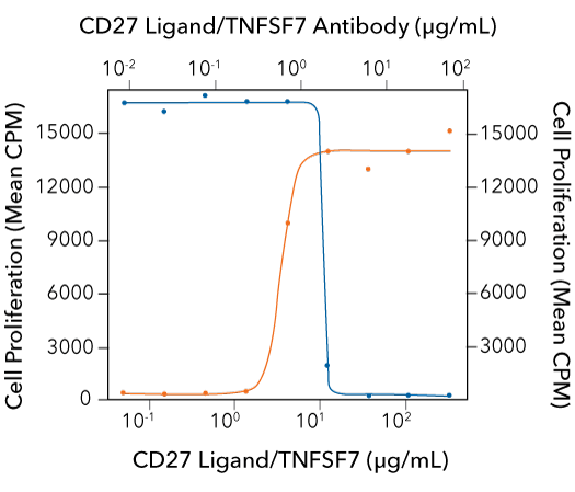
CD27 Ligand-induced Cell Proliferation is Neutralized Using a Rat Anti-Mouse CD27 Ligand Monoclonal Antibody. Mouse splenic T cells were treated with suboptimal amounts of a Hamster Anti-Mouse CD3 epsilon Monoclonal Antibody (R&D Systems, Catalog # MAB484) and increasing concentrations of Recombinant Mouse CD27 Ligand/TNFSF7 (R&D Systems, Catalog # 783-CL) and cell proliferation was assessed (orange line). The ED50 for this effect is 1.25-5 ug/mL in the presence of anti-CD3 epsilon. Under these conditions, proliferation stimulated by 10 ug/mL Recombinant Mouse CD27 Ligand/TNFSF7 was neutralized by treating the cells with increasing concentrations of a Rat Anti-Mouse CD27 Ligand/TNFSF7 Monoclonal Antibody (R&D Systems, Catalog # MAB783; blue line). The ND50 for this effect is typically 0.5-2 ug/mL.
Protein Characterization Using SEC-MALS Analysis

Recombinant Human TNF-alpha (HEK293-expressed) Protein SEC-MALS Recombinant human TNF-alpha (Catalog # 10291-TA) has a molecular weight (MW) of 52.3 kDa as analyzed by SEC-MALS, suggesting that this protein is a homotrimer. MW may differ from predicted MW due to post-translational modifications (PTMs) present (i.e. Glycosylation).
| SEC-MALS Data | Result |
| Retention Time | 18.6-19.0 min |
| MW-Predicted (Monomer) | 17.0 kDa |
| MW-MALS | 52.3 kDa |
| Polydispersity | 1 |
| System Suitability: BSA Monomer 66.4 ± 3.32 kDa | Pass |
Immunostaining of CD40 in Mouse Splenocytes Using R&D Systems Goat Anti-Mouse CD40 Polyclonal Antibody

Detection of CD40 in Mouse Splenocytes. Immersion-fixed mouse splenocytes were stained for CD40 expression using a Goat Anti-Mouse CD40/TNFRSF5 Antigen Affinity-purified Polyclonal Antibody (R&D Systems, Catalog # AF440). Cells were stained using a NorthernLights™ 557-conjugated Donkey Anti-Goat IgG Secondary Antibody (R&D Systems, Catalog # NL001; red) and the nuclei were counterstained with DAPI (Tocris, Catalog # 5748).
Cell Proliferation Induced by R&D Systems Recombinant Human CD40 Ligand and Neutralization by a Mouse Anti-Human CD40 Monoclonal Antibody

CD40 Ligand-induced Cell Proliferation is Neutralized Using a Mouse Anti-Human CD40 Monoclonal Antibody. In the presence of 20 ng/mL Recombinant Human IL-4 (R&D Systems, Catalog # 204-IL), human B cell-enriched peripheral blood mononuclear cells were treated with increasing concentrations of Recombinant Human CD40 Ligand/TNFSF5 (R&D Systems, Catalog # 6245-CL) and cell proliferation was assessed (orange line). The ED50 for this effect is 1-3 ug/mL in the presence of Recombinant Human IL-4. Under these conditions, proliferation stimulated by 10 ug/mL Recombinant Human CD40 Ligand/TNFSF5 was neutralized by treating the cells with increasing concentrations of a Mouse Anti-Human CD40/TNFRSF5 Monoclonal Antibody (R&D Systems, Catalog # MAB6322; blue line). At 5 ug/mL, this Mouse Anti-Human CD40 antibody will neutralize approximately 80% of Recombinant Human CD40 Ligand/TNFSF5-induced proliferation in the presence of 20 ng/mL Recombinant Human IL-4.
Apoptosis Induced by R&D Systems Recombinant Human Fas Ligand and Neutralization by a Goat Anti-Human Fas Ligand Polyclonal Antibody

Fas Ligand-induced Apoptosis is Neutralized Using a Goat Anti-Human Fas Ligand Polyclonal Antibody. In the presence of 10 ug/mL of a cross-linking Mouse Anti-Poly-Histidine Monoclonal Antibody (R&D Systems, Catalog # MAB050) Jurkat human acute T cell leukemia cells were treated with increasing concentrations of Recombinant Human Fas Ligand/TNFSF6 (R&D Systems, Catalog # 126-FL) and apoptosis was assessed (orange line). Under these conditions, the ED50 for this effect is 0.3-1.5 ng/mL. The apoptotic effect induced by 10 ng/mL Recombinant Human Fas Ligand/TNFSF6 was neutralized by treating the cells with increasing concentrations of a Goat Anti-Human Fas Ligand/TNFSF6 Antigen Affinity-purified Polyclonal Antibody (R&D Systems, Catalog # AF126; blue line). The ND50 for this effect is typically 0.012-0.072 ug/mL.
Detection of 4-1BB in Activated Mouse Splenocytes by Flow Cytometry

Detection of 4‑1BB/TNFRSF9/CD137 in Activated Mouse Splenocytes by Flow Cytometry. Activated mouse splenocytes were stained with an APC-conjugated Rat Anti-Mouse ICOS Monoclonal Antibody (R&D Systems, Catalog # FAB168A) and either (A) a PE-conjugated Rat Anti-Mouse 4-1BB Monoclonal Antibody (R&D Systems, Catalog # FAB937P) or (B) a PE-conjugated Rat IgG2A Isotype Control Antibody (R&D Systems, Catalog # IC006P).
Osteoclast-Like Cell Formation Induced by R&D Systems Recombinant Human TRANCE/RANK L and Neutralization by a Goat Anti-Human TRANCE Polyclonal Antibody
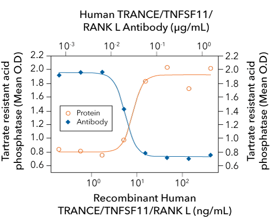
TRANCE/RANK Ligand-induced Osteoclast-Like Cell Formation is Neutralized Using a Goat Anti-Human TRANCE/RANK L Polyclonal Antibody. In the presence of 20 ng/mL Recombinant Mouse M-CSF (R&D Systems, Catalog # 416-ML), RAW264.7 mouse macrophages were treated with increasing concentrations of Recombinant Human TRANCE/TNFSF11/RANK L (R&D Systems, Catalog # 390-TN) and osteoclast-like cell formation was assessed using a tartrate-resistant acid phosphatase solution assay (orange line). Under these conditions, osteoclast-like cell formation elicited by 10 ng/mL Recombinant Human TRANCE/TNFSF11/RANK L was neutralized by treating the cells with increasing concentrations of a Goat Anti-Human TRANCE/TNFSF11/RANK L Antigen Affinity-purified Polyclonal Antibody (R&D Systems, Catalog # AF626; blue line). The ND50 is typically 0.01-0.04 ug/mL.
Featured TNF Superfamily Products


TNF Superfamily Blocking/Neutralizing Antibodies
TNF Superfamily Blocking/Neutralizing Antibodies
Blocking and neutralizing antibodies bind to their targets and either directly interfere with their functions or negatively regulate their downstream cellular effects. Bio-Techne offers a large selection of R&D Systems™ blocking or neutralizing antibodies against TNF superfamily ligands and receptors that have been validated to inhibit the activities of their target molecules in relevant biological assays.


Immunoassays for Detecting TNF Superfamily Ligands and Soluble Receptors
Immunoassays for Detecting TNF Superfamily Ligands and Soluble Receptors
From our complete, ready-to-use Quantikine™ ELISA Kits to our multiplex Luminex® Assays and fully automated Simple Plex™ Assays, you can count on our immunoassays to deliver accurate, reproducible, high-quality data for every experimental sample that you test.


Bulk Proteins
Bulk Proteins
If your experiments require large quantities of a particular protein, contact us for a bulk quote. We have the capacity and the expertise to scale up the production of any protein to meet your needs, and we offer economical pricing on bulk orders.
Featured TNF Superfamily Resources


TNFRSF Co-Stimulatory Immune Checkpoint Receptors
TNFRSF Co-Stimulatory Immune Checkpoint Receptors
Several members of the TNF receptor superfamily have been characterized as T cell and/or natural killer cell co-stimulatory receptors that enhance the survival, proliferation, and effector functions of these cells. As a result, researchers are investigating the effects of agonists of many of these receptors as potential therapeutic agents to improve anti-tumor immune responses.


TNF Superfamily Product Guide
TNF Superfamily Product Guide
Explore this guide to learn more about the different processes regulated by members of the TNF superfamily. This guide includes a complete listing of our TNF superfamily products and provides multiple data examples to demonstrate the testing our scientists have performed to ensure that our products will provide superior performance and lot-to-lot consistency.


Caspase Activation & Apoptosis Poster
Caspase Activation & Apoptosis Poster
Caspase activation and apoptosis occurs upon receipt of either an extrinsic or an intrinsic death signal. Use this poster to learn more about the molecules involved in mediating the two pathways of caspase activation, the different caspase activating complexes that can be formed, and the caspase domains and cleavage sites.

Custom Protein Services
If you can’t find the protein that you need on our website, contact us. From scratch protein development to customizing a protein from our catalog, our custom services team will create the protein that fits your experimental needs.