
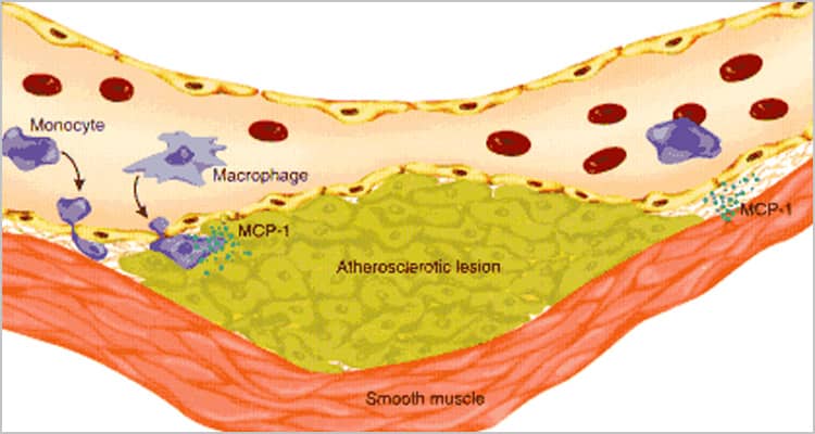
In response to mechanical stress, damage, or disease, vascular endothelial and smooth muscle cells upregulate many pro-inflammatory molecules. Chemokines promote the attraction of immune cells to the stressed area, adhesion proteins tether them in place, and cytokines and prostanoids promote their activation. Dysfunctional vascular cells also secrete proteases and morphogens to enhance tissue remodeling, and they upregulate cell surface proteins that reinforce their own inflammatory responses.
Chemokines
Activated vascular cells secrete chemokines that attract inflammatory immune cells expressing chemokine receptors. Chemokines of the CCL (-2/MCP-1, -3/MIP-1α, -4/MIP-1β, -5/RANTES, -8/MCP-2, -11/Eotaxin, -19/MIP-3β, -20/MIP-3α, -22/MDC) and CXCL (-1/KC, -2/MIP-2, -3/GROγ, -8/IL-8, -9/MIG, -10/IP-10, -16) families as well as CX3CL1/Fractalkine are inolved in this process.
Multianalyte Immunoassays
These assay platforms let you detect many proteins in one experiment. They are optimized to detect chemokines with high sensitivity, low background, and minimal cross-reactivity.
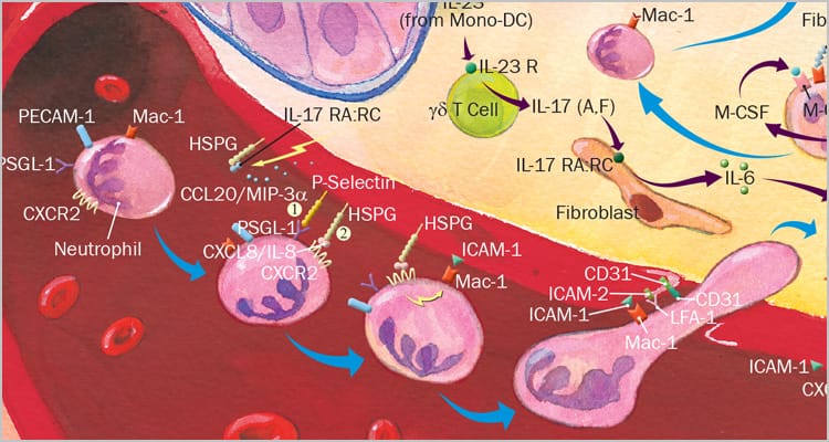
Adhesion
In order for circulating leukocytes to reach sites of tissue inflammation, they need to attach to regions of vascular endothelium near the infection or damaged tissue. Attachment is mediated by many adhesion proteins upregulated on activated endothelial cells, including Ig superfamily proteins (e.g. CD31, ICAM-1, VCAM-1), Integrins (e.g. αL/CD11a, αM/CD11b, αMβ2, αXβ2), and E-Selectin.
Multianalyte Immunoassays
These assay platforms let you detect many proteins in one experiment. They are optimized to detect analytes with high sensitivity, low background, and minimal cross-reactivity.
Cytokines
Activated vascular cells secrete multiple cytokines that regulate inflammation by contributing to the activation and differentiation of immune cells. In particular, T cell, macrophage, monocyte, dendritic cell, and neutrophil activation is highly responsive to vascular derived IFN-gamma, IL-6, -10, -17, -18, M-CSF, and TNF-alpha.

Multianalyte Immunoassays
These assay platforms let you detect many proteins in one experiment. They are optimized to detect cytokines with high sensitivity, low background, and minimal cross-reactivity.
Prostanoids
Vascular endothelial and smooth muscle cells release prostanoids under stress conditions. Among these inflammatory mediators, Prostaglandins PGD2 and PGE2 as well as Thromboxanes TXA2and TXB2 promote inflammation through interactions with their 7-TM receptors expressed on immune cells and the vasculature itself.

Morphogens
Tissue remodeling during angiogenesis, embryonic development, and wound healing involves matrix metalloproteases (e.g. MMP-1, -3, -9) to break down extracellular matrix and adhesive contacts, morphogenetic factors (e.g. BMP-2, -4, Noggin), to induce progenitor cell differentiation, and growth factors to promote cell proliferation (e.g. FGF basic, PDGF).
- MMP Bioactive Proteins
- MMP Antibodies and Conjugates
- MMP ELISA Kits
- Matrix Metalloprotease Inhibitors
- BMP-2 & -4 and Noggin Bioactive Proteins
- BMP-2 & -4 and Noggin Antibodies and Conjugates
- BMP-2 and BMP-4 ELISA Kits

Multianalyte Immunoassays
These assay platforms let you detect many proteins in one experiment. They are optimized to detect analytes with high sensitivity, low background, and minimal cross-reactivity.
Proinflammatory Receptors
Toll-like Receptors (TLRs), receptor for advanced glycation endproducts (RAGE/AGER), IL-6R alpha, CD40, and CD40 Ligand are expressed on vascular endothelial or smooth muscle cells. Their activation during inflammation or cell stress enhances the inflammatory response within the vasculature.

Multianalyte Immunoassays
These assay platforms let you detect many proteins in one experiment. They are optimized to detect analytes with high sensitivity, low background, and minimal cross-reactivity.
Additional Resources from R&DSystems, a Bio-Techne Brand
"Blood Brain Barrier Immune Cell Transmigration Pathway"
This interactive pathway covers leukocyte adhesion and BBB crossing in response to neural cell inflammation.
"Chemokine Signaling Pathway"
This pathway covers intracellular signaling downstream of C, CC, CXC, and CX3C chemokine receptors.
"Chemokine Superfamily Pathway"
This interactive pathway sorts out the binding specifities of chemokines for their receptors.
"Immunology Pathways"
This collection of pathways covers intracellular signaling downstream of many cytokine receptors.
"Pathogen or Damage-activated C-Type Lectin Receptor Signaling Pathway"
This interactive pathway covers intracellular signaling in response to infection or tissue damage.
"Toll-Like Receptor Signaling Pathway"
This interactive pathway covers intracellular TLR signaling in response to microbial infection.
"Atherosclerosis Disease Progression Poster"
This poster covers healthy vascular function, endothelial damage, plaque progression, and plaque rupture.
"Atherosclerosis Disease Progression Cell Markers"
Interactive tool that covers secreted, cell surface, and intracellular molecules in vascular and immune cells.
"Chemokine Nomenclature Resource"
This reference organizes the many alternative chemokine names into the systemic nomenclature.
"The Chemokine Superfamily: Critical Regulators of Homeostasis & Inflammation Poster"
This poster covers chemokine function and signaling plus receptor expression on immune cells.
"Periodic Table of Human Cytokine and Chemokine Families Poster"
This poster presents biochemical and receptor binding information for cytokines and chemokines.
"Periodic Table of Human Developmental Factors Poster"
This poster presents biochemical and receptor binding information for developmental and morphogenic factors.
"Unit Conversion Table"
This table correlates the specific activity of R&D Systems® growth factors with World Health Organization standards.
"ProteinSimple® Analytical Instrumentation"
Meet our protein characterization problem solvers – Simple Western™, Maurice, Simple Plex™, Single-Cell Western, and MFI™Systems.
"RNAscope® and BaseScope™ Assays"
Revolutionary ISH assays capture gene expression in situ at single cell level in intact tissue.
"Bioprocessing: From Start to Finish"
Instruments, GMP reagents, and services for clone selection through final product characterization.
"Cell and Gene Therapy Solutions"
Pioneering tools to simplify your workflow – cell separation, cell culture, and quality control.
"GMP Grade Proteins"
Cytokines and growth factors for ancillary use in preclinical to clinical studies.